Deep Venous Disease (DVD)
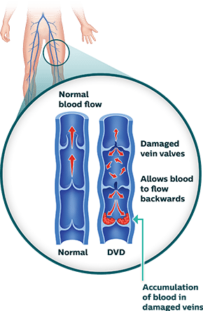
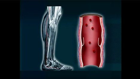
Veins are responsible for returning blood to the heart. But when they become damaged, and are unable to return blood effectively, they can cause complications throughout the body, including increased pressure on vessel walls and pooling of blood in the legs.1 Several types of deep venous disease (DVD) conditions exist including venous compression, deep venous obstruction (DVO), deep venous insufficiency (DVI), nonthrombotic iliac vein lesions (NIVL), popliteal entrapment syndrome, and thoracic outlet syndrome.2 Each condition leads to the compression of veins in different parts of the body and can be either acute or chronic. Over time, these conditions can lead to chronic swelling and various vein problems and can put you at risk for deep vein thrombosis (DVT). Learn more about the deep venous disease symptoms and how DVD can be treated.
Symptoms of DVD
Because the symptoms of both DVI and DVO are similar, the following sections will refer to both vein malfunctions.
DVI
")
DVI is a malfunctioning of the vein valves making it difficult for the veins to send blood back to the heart.3
DVO
DVO is a compression of the vein resulting in either a partial or complete obstruction of the blood flow.
Sometimes people who may be otherwise healthy—most notably young women—may begin to experience various symptoms.4 For others with conditions such as NIVL, patients may not experience symptoms until triggered by an event such as an injury. DVD symptoms include:
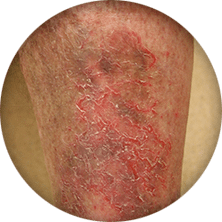
Skin Changes
Color changes in the leg including: paleness or blueness, skin rash, redness and itching. Advanced DVD can exhibit skin ulcerations.

Pain
Pain, deep throbbing diffuse pain in the leg muscles that occur after prolonged standing.
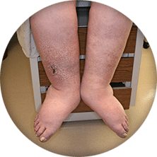
Swelling
Deep venous disease (DVD) may cause uncomfortable swelling in the legs, feet and ankles.
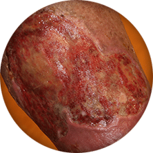
Non-healing Wounds
Sores or wounds on the legs, located above the ankle, that won’t heal or aren’t healing well.
Non-healing wound image courtesy of Dr. Raghu Kolluri.
DVT Diagnosis and Treatment
Problems deep in the veins—DVO or DVI—can signal several serious conditions, which is why it is important to obtain a diagnosis right away. A doctor may use a combination of physical exam, medical history and one of several tools to make a diagnosis and determine if it is DVO or DVI.5 Deep venous obstruction (DVO) is either a compression of the vein limiting the flow of blood, or a complete blockage stopping the flow of blood. Deep venous insufficiency (DVI) is a condition where the flow of blood through the veins is inadequate, usually caused by malfunctioning valves in the vein.
Deep Venous Obstruction (DVO) Diagnostic Procedures
Photos of Diagnostic Procedures
")
Duplex Ultrasound
A tool that uses sound waves to look at the flow of blood in the veins.
")
Magnetic Resonance Venography (MRV) or Computed Tomography Venography
A type of imaging tool that uses magnetic wave and radio energy to locate and measure the severity of a blocked blood vessel.
")
IVUS
A special imaging device that uses a small catheter to image the interior of blood vessels and to help identify deep venous compression and its source.
Videos of Diagnostic Procedures
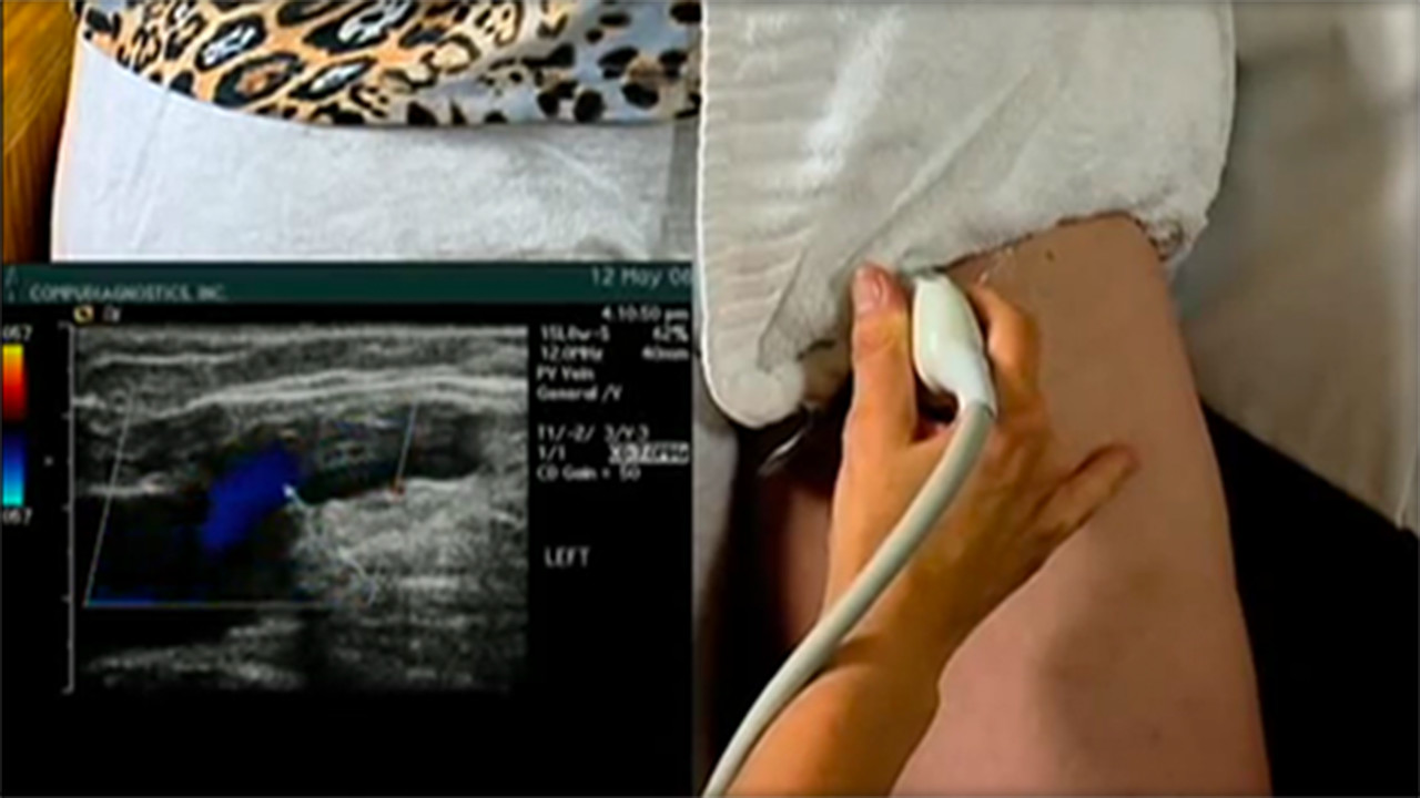
Duplex Ultrasound
This is a non-invasive test which uses sound waves to create a color map of the blood vessels and examines blood flow in your legs.

Magnetic Resonance Venography (MRV) or Computed Tomography Venography
A type of imaging tool that uses magnetic wave and radio energy to locate and measure the severity of a blocked blood vessel.
DVT Treatments
Despite serious complications, DVT can be treated if detected early. Certain treatments may work for some patients, but not others. A vascular specialist may recommend treatments that prevent the blood clot from enlarging and moving to the lungs or other therapies to dissolve the clot right away. These treatments may include:3 Photos of DVT Treatments
")
Medication
Some medications can be taken orally while some need to be injected. They are used to reduce the chances of a blood clot and facilitate blood flow.
")
Angioplasty / Stenting
This procedure involves the use of a catheter to open narrowed or blocked veins, increasing blood flow. It may also involve the placement of a stent to maintain the diameter of the blood vessel.
Deep Venous Insufficiency (DVI) Diagnostic Procedures
Deep venous insufficiency can encompass a blockage of a vein upstream or downstream resulting in an insufficiency of flow. Photos of Diagnostic Procedures
")
Duplex Ultrasound
This is a non-invasive test which uses sound waves to create a color map of the blood vessels and examines blood flow in your legs.
DVI Treatments
Photos of DVI Treatments
")
Compression Stockings
These are recommended not only to prevent swelling, but to relieve pain associated with venous compression.
Videos of DVI Treatments
Compression Stockings
These are recommended not only to prevent swelling, but to relieve pain associated with venous compression.
DVD Patient Stories
Deep venous insufficiency can cause pooling of blood in the legs and feet causing swelling and pain especially after prolonged standing. Take the step and learn from other patients what the road to recovery is like.

I felt pain in my leg all day, like I had pulled a muscle. That evening, I collapsed.”

More Patient Stories
PAD Stories

Many PAD patients experience a gradual decline in activity level and quality of life which can go unnoticed for years. Learn about Joe’s suspicions and his perseverance to seek help.
CLI Stories

Early intervention can be key for critical limb ischemia (CLI). Learn about Mark’s struggles and triumph to overcome CLI.
DVT Stories

Deep vein thrombosis (DVT) can strike quickly with little notice. Learn more about Reid’s condition and his road to recovery.
Helpful Resources

PVD Doctor
Discussion Guide
Get helpful tips and advice on how to talk to your doctor about a PVD screening.

DVD Brochure
Find out more information on PAD by downloading this helpful brochure.
Third-Party Resources
Helpful Websites
Blogs
Apps
A mobile app built by cardiologists, to simplify understanding of most cardiac and peripheral vascular conditions and treatments.
CardioVisual
")
")
")
Find a Doctor that Treats DVD
Consult your doctor to learn more about DVD risks and how to maintain your vascular health. You can also use our tool below to find a specialist near you.
This tool is not inclusive of all specialists. Consult with your insurance provider to find specialists that are covered within your network.
Are You a Healthcare Professional?
Find out how to register your practice with us.
1. Henke, P. “Chronic Venous Insufficiency.” Society for Vascular Surgery. n.d. 2. Butros et al. “Venous compression syndromes: clinical features, imaging findings and management.” Br J Radiol. 2013 Oct; 86(1030): 20130284. 3. “Venous insufficiency.” MedlinePLUS. U.S. National Library of Medicine. June 6, 2016. 4. Raju et al. “High prevalence of nonthrombotic iliac vein lesions in chronic venous disease: A permissive role in pathogenicity.” Journal of Vascular Surgery. Volume 44, Issue 1, Pages 136–144 5. “Venous Thromboembolism (Blood Clots).” Centers for Disease Control and Prevention. August 13, 2015
Disclaimer
The opinions and clinical experiences presented herein are for informational purposes only. Individual results may vary depending on a variety of patient-specific attributes and related factors. Dr. Raghu Kolluri has been compensated by Philips for his services in preparing and providing this material for Philips further use and distribution. We offer links to third-party websites that may be of interest to our website visitors and in no way, represent any affiliation or endorsement of the information provided on those linked websites. These links are provided solely for your convenience and to assist you in learning more information on this topic. However, Philips does not control, endorse or guarantee the accuracy of the information contained on the linked sites. In addition, Philips makes no representations or warranties of any kind with regards to any third-party websites or information contained therein. If you have any questions or concerns about the information on the linked third-party websites, please contact the third-party websites directly.